Price includes epidural, and follow-up appointments at 2, 4, 6, and 8 weeks.
Introduction to Intervertebral Disc Disease (IVDD)
IVDD in dogs is a common cause of back pain, weakness, and the inability to walk or feel the front and/or back legs. IVDD can affect any part of the canine spine, however, it more commonly affects the thoracolumbar (‘mid-back’) spine.

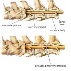
Canine Spine with IVDD. Reprinted with permission by the copyright owner, Hill’s Pet Nutrition, Inc. Fig 1 + 2

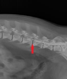
Myelogram of the thoracolumbar spine showing herniated intervertebral disc (arrow) Fig 3
Symptoms & Signs of IVDD
Most commonly dogs herniate (or ‘slip’) a disc (Figure 2), which may forcibly strike and/or compress the spinal cord.
This leads to signs of IVDD. Signs of thoracolumbar IVDD include back pain, pelvic limb ataxia (walking wobbly), inability to stand, inability to move the rear legs, or even inability to feel the back legs. Fecal or urinary incontinence can also occur in severe cases. A grading scale is used in dogs:
- Grade 1) Pain Only – These dogs can walk normally, but exhibit signs of pain including reluctance to move, reluctance to jump, shivering, crying, muscle spasms, and/or a tense abdomen.
- Grade 2) Ambulatory Paraparesis – These dogs can walk but are weak and wobbly in the rear legs. They may cross their back legs when walking, splay out, knuckle over, or stumble on their back legs.
- Grade 3) Non-Ambulatory Paraparesis – These dogs are still able to move their legs and wag their tails but are not strong enough to support their own weight and walk.
- Grade 4) Paraplegia – These dogs have no voluntary movement in the rear legs.
- Grade 5) Paraplegia with Absent Nociception (no ‘deep pain’) – In addition to being unable to move the back legs, they are unable to feel their back legs.
Diagnosis of IVDD
There are other diseases that can cause similar clinical signs of spinal cord disease including meningitis/myelitis, spinal tumors, trauma, infection, malformations, and vascular injury, among other problems.
- Neurological Examination – Gives the veterinarian an idea of which of these are more likely than others, but tests are necessary to accurately determine the cause.
- Spinal Radiographs – Useful for screening for disc infection and bony tumors, but typically are insufficient to diagnose IVDD.
- Myleography – Involves injecting contrast around the spinal cord to visualize it on radiographs. (Figure 3)
- Computed Tomography (CT) Scan – Useful (often combined with a myelogram) and allows the body to be visualized in ‘slices.’
- Magnetic Resonance Imaging (MRI) – Considered the imaging modality of choice when visualizing the soft tissues of the body, including the nervous system. High-field MRI offers several advantages over low-field MRI, CT, and/or myelography.
Treatment of IVDD
Treatment depends on the severity of the signs, but can be divided into two options:
- Non-Surgical – For dogs with a first episode of back pain or mild pelvic limb ataxia, a conservative approach of cage rest and medications may be elected.
- Surgical – Dogs with more severe signs (grade two through five) or recurrent or persistent back pain that does not respond to rest and medications are best managed with surgery to decompress the spinal cord.
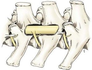
After examination and advanced imaging, the disc herniation can be located. A hemilaminectomy (removal of one side of the vertebra) can be performed to allow the surgeon to remove the herniated disc material and decompress the spinal cord.

Hemilaminectomy Fig 4
Outcome
Postoperative complications can include:
- Myelogram can cause seizures in the first 24 hours after the procedure (very rare)
- Incisional infection
- Thromboembolism
- Continued wobbly walk or dragging hind toes when walking
- Many patients have another disc herniated later in life
Prognosis varies based on the grade and location of disease. The lower the grade, the better the prognosis. Generally, the sooner the issue is addressed, the better the overall prognosis.
Left untreated, a Grade 1 can progress to more serious problems. Severe disc ruptures can lead to permanent spinal cord injury.